

Alexandros Sfakianakis
Anapafseos 5 . Agios Nikolaos
Crete.Greece.72100
2841026182
Anapafseos 5 . Agios Nikolaos
Crete.Greece.72100
2841026182
6948891480
Verification of soman-related nerve agents via detection of phosphonylated adducts from rabbit albumin in vitro and in vivoAbstractA major challenge in organophosphate compound (OP) and OP nerve agent (OPNA) research has been in the identification and utilization of reliable biomarkers for rapid, sensitive, and efficient detection of OP exposure. Albumin has been widely studied as a biomarker for retrospective verification of exposure to OPNAs, including soman (GD), by detecting the phosphonylation of specific amino acid residues. The aim of the present study was to identify binding sites between GD and rabbit serum albumin in vitro and in vivo. A nano-liquid chromatography coupled with a quadrupole-orbitrap mass spectrometry (nLC-Q-Orbitrap-MS) was used to examine the GD-modified adducts of rabbit albumin. A total of 11 GD-modified sites were found in rabbit serum albumin across three experimental models. The following five GD-modified rabbit albumin sites, which were all lysine residues, were established in vivo: K188, K329, K162, K233, and K525. Two of these five lysine residues, K188 in peptide EK*ALISAAQER and K162 in peptide YK*AILTECCEAADK, were stable for at least 7 days in vivo. Molecular simulation of the GD–albumin interaction provided theoretical evidence for reactivity of the identified lysine residues. The findings suggest that these modifiable lysine residues are potential biomarkers of GD exposure for retrospective analysis by Q-Orbitrap-MS. |
| Comment on 'Kim, S.-J., Choi, E.-J., Choi, G.-W., Lee, Y.-B., and Cho, H.-Y. (2019). Exploring sex differences in human health risk assessment for PFNA and PFDA using a PBPK model, Arch Toxicol 93:311–330' |
Uptake and effects of orally ingested polystyrene microplastic particles in vitro and in vivoAbstractEvidence exists that humans are exposed to plastic microparticles via diet. Data on intestinal particle uptake and health-related effects resulting from microplastic exposure are scarce. Aim of the study was to analyze the uptake and effects of microplastic particles in human in vitro systems and in rodents in vivo. The gastrointestinal uptake of microplastics was studied in vitro using the human intestinal epithelial cell line Caco-2 and thereof-derived co-cultures mimicking intestinal M-cells and goblet cells. Different sizes of spherical fluorescent polystyrene (PS) particles (1, 4 and 10 µm) were used to study particle uptake and transport. A 28-days in vivo feeding study was conducted to analyze transport at the intestinal epithelium and oxidative stress response as a potential consequence of microplastic exposure. Male reporter gene mice were treated three times per week by oral gavage with a mixture of 1 µm (4.55 × 10⁷ particles), 4 µm (4.55 × 10⁷ particles) and 10 µm (1.49 × 10⁶ particles) microplastics at a volume of 10 mL/kg/bw. Effects of particles on macrophage polarization were investigated using the human cell line THP-1 to detect a possible impact on intestinal immune cells. Altogether, the results of the study demonstrate the cellular uptake of a minor fraction of particles. In vivo data show the absence of histologically detectable lesions and inflammatory responses. The particles did not interfere with the differentiation and activation of the human macrophage model. The present results suggest that oral exposure to PS microplastic particles under the chosen experimental conditions does not pose relevant acute health risks to mammals. |
Proteomic analysis of hippocampal proteins in acrylamide-exposed Wistar ratsAbstractAcrylamide has been used industrially and also found in certain foods cooked at high temperatures. Previous reports described acrylamide-related human intoxication who presented with ataxia, memory impairment, and/or illusion. The aim of this study was to characterize the molecular mechanisms of neurotoxicity of acrylamide by analyzing the expression levels of various proteins in the hippocampus of rats exposed to acrylamide. Male Wistar rats were administered acrylamide by gavage at 0, 2, and 20 mg/kg for 1 week or 0, 0.2, 2, and 20 mg/kg for 5 weeks. At the end of the experiment, the hippocampus was dissected out and proteins were extracted for two-dimensional difference gel electrophoresis combined with matrix-assisted laser-desorption ionization time-of-flight/time-of-flight mass spectrometry (MALDI-TOF/TOF/MS). MALDI-TOF/TOF/MS identified significant changes in two proteins in the 1-week and 22 proteins in the 5-week exposure groups. These changes were up-regulation in 9 and down-regulation in 13 proteins in the hippocampus of rats exposed to acrylamide at 20 mg/kg for 5 weeks. PANTHER overrepresentation test based on the GO of biological process showed significant overrepresentation in proteins annotated to nicotinamide nucleotide metabolic process, coenzyme biosynthetic process, pyruvate metabolic process, and carbohydrate metabolic process. The test also showed significant overrepresentation in proteins annotated to creatinine kinase activity for the GO of molecular function as well as myelin sheath, cytoplasmic part, and cell body for the GO of cellular component. Comparison with a previous proteomic study on hippocampal proteins in rats exposed to 1-bromopropane identified triosephosphate isomerase, mitochondrial creatine kinase U-type, creatine kinase β-type and proteasome subunit α type-1 as proteins affected by exposure to acrylamide and 1-bromopropane, suggesting a common mechanism of neurotoxicity for soft electrophiles. |
Ecdysteroids as non-conventional anabolic agent: performance enhancement by ecdysterone supplementation in humansAbstractRecent studies suggest that the anabolic effect of ecdysterone, a naturally occurring steroid hormone claimed to enhance physical performance, is mediated by estrogen receptor (ER) binding. In comparison with the prohibited anabolic agents (e.g., metandienone and others), ecdysterone revealed to be even more effective in a recent study performed in rats. However, scientific studies in humans are very rarely accessible. Thus, our project aimed at investigating the effects of ecdysterone-containing products on human sport exercise. A 10-week intervention study of strength training of young men (n = 46) was carried out. Different doses of ecdysterone-containing supplements have been administered during the study to evaluate the performance-enhancing effect. Analysis of blood and urine samples for ecdysterone and potential biomarkers of performance enhancement has been conducted. To ensure the specificity of the effects measured, a comprehensive screening for prohibited performance-enhancing substances was also carried out. Furthermore, the administered supplement has been tested for the absence of anabolic steroid contaminations prior to administration. Significantly higher increases in muscle mass were observed in those participants that were dosed with ecdysterone. The same hypertrophic effects were also detected in vitro in C2C12 myotubes. Even more relevant with respect to sports performance, significantly more pronounced increases in one-repetition bench press performance were observed. No increase in biomarkers for liver or kidney toxicity was noticed. These data underline the effectivity of an ecdysterone supplementation with respect to sports performance. Our results strongly suggest the inclusion of ecdysterone in the list of prohibited substances and methods in sports in class S1.2 "other anabolic agents". |
Neurotoxicity of Micrurus lemniscatus lemniscatus (South American coralsnake) venom in vertebrate neuromuscular preparations in vitro and neutralization by antivenomAbstractWe investigated the effect of South American coralsnake (Micrurus lemniscatus lemniscatus) venom on neurotransmission in vertebrate nerve–muscle preparations in vitro. The venom (0.1–30 µg/ml) showed calcium-dependent PLA2 activity and caused irreversible neuromuscular blockade in chick biventer cervicis (BC) and mouse phrenic nerve–diaphragm (PND) preparations. In BC preparations, contractures to exogenous acetylcholine and carbachol (CCh), but not KCl, were abolished by venom concentrations ≥ 0.3 µg/ml; in PND preparations, the amplitude of the tetanic response was progressively attenuated, but with little tetanic fade. In low Ca2+ physiological solution, venom (10 µg/ml) caused neuromuscular blockade in PND preparations within ~ 10 min that was reversible by washing; the addition of Ca2+ immediately after the blockade temporarily restored the twitch responses, but did not prevent the progression to irreversible blockade. Venom (10 µg/ml) did not depolarize diaphragm muscle, prevent depolarization by CCh, or cause muscle contracture or histological damage. Venom (3 µg/ml) had a biphasic effect on the frequency of miniature end-plate potentials, but did not affect their amplitude; there was a progressive decrease in the amplitude of evoked end-plate potentials. The amplitude of compound action potentials in mouse sciatic nerve was unaffected by venom (10 µg/ml). Pre-incubation of venom with coralsnake antivenom (Instituto Butantan) at the recommended antivenom:venom ratio did not neutralize the neuromuscular blockade in PND preparations, but total neutralization was achieved with a tenfold greater volume of antivenom. The addition of antivenom after 50% and 80% blockade restored the twitch responses. These results show that M. lemniscatus lemniscatus venom causes potent, irreversible neuromuscular blockade, without myonecrosis. This blockade is apparently mediated by pre- and postsynaptic neurotoxins and can be reversed by coralsnake antivenom. |
The in vivo developmental toxicity of diethylstilbestrol (DES) in rat evaluated by an alternative testing strategyAbstractIn the present study, we evaluated an alternative testing strategy to quantitatively predict the in vivo developmental toxicity of the synthetic hormone diethylstilbestrol (DES). To this end, a physiologically based kinetic (PBK) model was defined that was subsequently used to translate concentration–response data for the in vitro developmental toxicity of DES, obtained in the ES-D3 cell differentiation assay, into predicted in vivo dose–response data for developmental toxicity. The previous studies showed that the PBK model-facilitated reverse dosimetry approach is a useful approach to quantitatively predict the developmental toxicity of several developmental toxins. The results obtained in the present study show that the PBK model adequately predicted DES blood concentrations in rats. Further studies revealed that DES tested positive in the ES-D3 differentiation assay and that DES-induced inhibition of the ES-D3 cell differentiation could be counteracted by the estrogen receptor alpha (ERα) antagonist fulvestrant, indicating that the in vitro ES-D3 cell differentiation assay was able to mimic the role of ERα reported in the mode of action underlying the developmental toxicity of DES in vivo. In spite of this, combining these in vitro data with the PBK model did not adequately predict the in vivo developmental toxicity of DES in a quantitative way. It is concluded that although the EST qualifies DES as a developmental toxin and detects the role of ERα in this process, the ES-D3 cell differentiation assay of the EST apparently does not adequately capture the processes underlying DES-induced developmental toxicity in vivo. |
Novel insight in estrogen homeostasis and bioactivity in the ACI rat model of estrogen-induced mammary gland carcinogenesisAbstractDespite being widely used to investigate 17β-estradiol (E2)-induced mammary gland (MG) carcinogenesis and prevention thereof, estrogen homeostasis and its significance in the female August Copenhagen Irish (ACI) rat model is unknown. Thus, levels of 12 estrogens including metabolites and conjugates were determined mass spectrometrically in 38 plasmas and 52 tissues exhibiting phenotypes ranging from normal to palpable tumor derived from a representative ACI study using two different diets. In tissues, 40 transcripts encoding proteins involved in estrogen (biotrans)formation, ESR1-mediated signaling, proliferation and oxidative stress were analyzed (TaqMan PCR). Influence of histo(patho)logic phenotypes and diet on estrogen and transcript levels was analyzed by 2-way ANOVA and explanatory variables influencing levels and bioactivity of estrogens in tissues were identified by multiple linear regression models. Estrogen profiles in tissue and plasma and the influence of Hsd17b1 levels on intra-tissue levels of E2 and E1 conclusively indicated intra-mammary formation of E2 in ACI tumors by HSD17B1-mediated conversion of E1. Proliferation in ACI tumors was influenced by Egfr, Igf1r, Hgf and Met levels. 2-MeO-E1, the only oxidative estrogen metabolite detected above 28–42 fmol/g, was predominately observed in hyperplastic tissues and intra-tissue conversion of E1 seemed to contribute to its levels. The association of the occurrence of 2-MeO-E1 with higher levels of oxidative stress observed in hyperplastic and tumor tissues remained equivocal. Thus, the present study provides mechanistic explanation for previous and future results observed in the ACI model. |
Okadaic acid activates Wnt/β-catenin-signaling in human HepaRG cellsAbstractThe lipophilic phycotoxin okadaic acid (OA) occurs in the fatty tissue and hepatopancreas of filter-feeding shellfish. The compound provokes the diarrhetic shellfish poisoning (DSP) syndrome after intake of seafood contaminated with high levels of the DSP toxin. In animal experiments, long-term exposure to OA is associated with an elevated risk for tumor formation in different organs including the liver. Although OA is a known inhibitor of the serine/threonine protein phosphatase 2A, the mechanisms behind OA-induced carcinogenesis are not fully understood. Here, we investigated the influence of OA on the β-catenin-dependent Wnt-signaling pathway, addressing a major oncogenic pathway relevant for tumor development. We analyzed OA-mediated effects on β-catenin and its biological function, cellular localization, post-translational modifications, and target gene expression in human HepaRG hepatocarcinoma cells treated with non-cytotoxic concentrations up to 50 nM. We detected concentration- and time-dependent effects of OA on the phosphorylation state, cellular redistribution as well as on the amount of transcriptionally active β-catenin. These findings were confirmed by quantitative live-cell imaging of U2OS cells stably expressing a green fluorescent chromobody which specifically recognize hypophosphorylated β-catenin. Finally, we demonstrated that nuclear translocation of β-catenin mediated by non-cytotoxic OA concentrations results in an upregulation of Wnt-target genes. In conclusion, our results show a significant induction of the canonical Wnt/β-catenin-signaling pathway by OA in human liver cells. Our data contribute to a better understanding of the molecular mechanisms underlying OA-induced carcinogenesis. |
The Toll-like receptor agonist imiquimod is metabolized by aryl hydrocarbon receptor-regulated cytochrome P450 enzymes in human keratinocytes and mouse liverAbstractThe Toll-like receptor 7 agonist imiquimod (IMQ) is an approved drug for the topical treatment of various skin diseases that, in addition, is currently tested in multiple clinical trials for the immunotherapy of various types of cancers. As all of these trials include application of IMQ to the skin and evidence exists that exposure to environmental pollutants, i.e., tobacco smoke, affects its therapeutic efficacy, the current study aims to elucidate the cutaneous metabolism of the drug. Treatment of human keratinocytes with 2.5 µM benzo[a]pyrene (BaP), a tobacco smoke constituent and aryl hydrocarbon receptor (AHR) agonist, for 24 h induced cytochrome P450 (CYP) 1A enzyme activity. The addition of IMQ 30 min prior measurement resulted in a dose-dependent inhibition of CYP1A activity, indicating that IMQ is either a substrate or inhibitor of CYP1A isoforms. Incubation of 21 recombinant human CYP enzymes with 0.5 µM IMQ and subsequent LC–MS analyses, in fact, identified CYP1A1 and CYP1A2 as being predominantly responsible for IMQ metabolism. Accordingly, treatment of keratinocytes with BaP accelerated IMQ clearance and the associated formation of monohydroxylated IMQ metabolites. A co-incubation with 5 µM 7-hydroxyflavone, a potent inhibitor of human CYP1A isoforms, abolished basal as well as BaP-induced IMQ metabolism. Further studies with hepatic microsomes from CD-1 as well as solvent- and β-naphthoflavone-treated CYP1A1/CYP1A2 double knock-out and respective control mice confirmed the critical contribution of CYP1A isoforms to IMQ metabolism. Hence, an exposure to life style-related, dietary, and environmental AHR ligands may affect the pharmacokinetics and, thus, treatment efficacy of IMQ. |
| Early-age Ndufs4 knockout mice are an inappropriate animal model of Leigh syndrome |
| Early detection of elevated lactate levels in a mitochondrial disease model using chemical exchange saturation transfer (CEST) and magnetic resonance spectroscopy (MRS) with 7T MR imaging |
Proposal of a new method to prove that unnecessary information is not drawn on the image using statistical analysisAbstractThe purpose of this study is to propose a new method of image evaluation using statistical analysis. We used the Sign test and the Wilcoxon test to analyze the statistical significance of image differences. Using this method, we evaluated whether the small electrode of the DAP meter appears in the X-ray image. Two observed values, which were obtained by averaging all values under all exposure conditions, were compared. All the observation tests showed the same sign. Thus, the results proved that the small electrode of the DAP meter is not present on the image. Using this method, it became possible to prove that the electrode was not depicted, which was impossible to determine using conventional methods. The method combining both the Sign test and the Wilcoxon test can be useful in image evaluation. |
Development of a new image manipulation system based on detection of electroencephalogram signals from the operator's brain: a feasibility studyAbstractPhysicians require an adequate display system with a console within arm's reach to view images during surgical operations and interventional radiological examinations. However, manipulation of the console by physicians themselves may not be possible because their hands may be otherwise engaged. In this study, an image manipulation system using an electroencephalogram (EEG) sensor mounted on the operator's head was developed. In this system, data acquired by the device is used to manipulate images, and the output can be converted to commands for various actions such as paging, which can be controlled by the operator's eye-blink, and zooming of a region indicated by the cursor, which can be controlled by the operator's mental concentration. In this study, the MindWave Mobile headset was used as EEG sensor, and AZEWIN for the display system. Ten observers were enrolled and fitted with EEG device to determine the threshold values of blink strength and attention; threshold value of 100 for blink strength and 65 for attention were determined. Thirty-one observers were enrolled and fitted with EEG device to investigate average response-time; the average response time for detecting paging was 0.43 ± 0.02 s, and that for zooming was 5.85 ± 0.56 s. Thus, the proposed image manipulation system using the operator's EEG signals enabled physicians to assess and manipulate images without using their hands. |
Voxel-based morphometry analysis of double inversion-recovery magnetic resonance imaging for detecting microscopic lesions: a simulation studyAbstractDouble inversion-recovery (DIR) imaging has the potential to improve the detection of subcortical lesions through the use of voxel-based morphometry (VBM) analysis. The aim of this study was to clarify the characteristics of detectable lesions by performing a VBM analysis on DIR images of simulated lesions. Twenty healthy volunteers underwent magnetic resonance imaging using a head three-dimensional DIR sequence. The images were processed using SPM12; then, the selected images with simulated lesions were analyzed via VBM. The VBM results were evaluated using free-response receiver-operating characteristic curves and a receiver-operating characteristic analysis. The sensitivity was 100% (5/5), with 5.6 false-positive objects per case, in simulated lesions with a contrast of 0.6 and a size of 2.4 mm. The sensitivity was 80% (4/5), with 5.4 false-positive objects per case, in simulated lesions with a contrast of 0.5 and a size of 2.4 mm. The mean area under the curve value was increased from 0.783 to 0.883 using VBM, with a statistically significant difference (p < 0.01). The VBM analysis of the DIR images using SPM alone showed the potential to detect subcortical microscopic lesions. Early detection of Alzheimer's disease may be possible by adapting VBM in the clinical setting. |
Estimation and validation of the frequency responses of a scanner system and an image reconstruction system in X-ray computed tomographyAbstractIn computed tomography, factors that theoretically affect the modulation transfer function (MTF) in the region near the isocenter are the frequency responses of the scanner system (MTFS) and reconstruction processing (MTFA). Although MTFS and MTFA are performance indices that are not disclosed to the users, both can be estimated by the measured MTF with the use of theoretical formulas. In this study, we proposed two methods to obtain the MTFS and MTFA, and confirm their validity. The first method to obtain the MTFS and MTFA uses a theoretical formula and the measured MTF. Another method uses the measured MTF and the noise power spectrum. In both the methods, the MTFS and MTFA were obtained separately. By our proposed methods, performance indices that are not usually disclosed to the users can be known. |
Appropriate echo time selection for quantitative susceptibility mappingAbstractThe purpose of our study was to clarify the dependence of quantitative susceptibility mapping (QSM) on echo time (TE). We constructed a phantom consisting of six tubes; three tubes were filled with different concentrations (0.5, 1.0, and 2.5 mM) of gadopentetate dimeglumine (Gd-DTPA), and three were filled with different concentrations (100, 200, and 350 mg/mL) of calcium hydroxyapatite. Real and imaginary images from multi-echo spoiled gradient-echo data (12 echoes) were acquired. We then used four datasets with three serial echoes. The QSM procedure consists of four steps: field map estimation, phase unwrapping, background removal, and dipole inversion. For each sample, we compared the measured mean susceptibility value with the theoretical susceptibility value and conducted a linear regression analysis. Accordingly, the relationship between the measured susceptibility and concentration of Gd-DTPA was shown to agree well with the theoretical values (TEs = 16.4, 20.8, and 25.2 ms; slope = 0.24, R2 = 1.00). Furthermore, the relationship between the measured susceptibility and concentration of hydroxyapatite also showed good linearity (TEs = 16.4, 20.8, and 25.2 ms; slope = − 0.00121, R2 = 1.00). In conclusion, the optimization of the TE in QSM makes it possible to obtain more detailed information regarding the susceptibility of biomaterials. |
Clinical application of biological fingerprints extracted from averaged chest radiographs and template-matching technique for preventing left–right flipping mistakes in chest radiographyAbstractWe aimed to evaluate the identification performance achieved using biological fingerprints extracted from averaged chest radiographs and template-matching techniques for the prevention of left–right flipping mistakes. We produced averaged chest radiographs for each sex by averaging 100 posteroanterior chest radiographs. Further, 400 and 566 chest radiographs were used in consistency and validation tests, respectively, and they were flipped horizontally to produce flipped chest radiographs under the assumption that the left–right flipping mistake occurred. The correlation values obtained with chest radiographs and those obtained with flipped chest radiographs were calculated. When we used correlation indices calculated from the correlation values from four biological fingerprints except for the lung apex, 96.5% (386/400) and 95.8% (542/566) of the left or right sides were identified correctly in the consistency and validation tests, respectively. This result indicates that our proposed method would be promising for the prevention of left–right flipping mistakes. |
A new approach for detecting abnormalities in mammograms using a computer-aided windowing system based on Otsu's methodAbstractBreast cancer is the most common cancer and the leading cause of cancer deaths in women worldwide. This study aimed to provide an automatic windowing method in mammograms, based on the principles of Otsu's thresholding function, to help radiologists more easily detect abnormalities on mammograms. A total of 322 mammographic images from the Mammographic Image Analysis Society (MIAS) database were used in the present study. The image background was removed based on Otsu's method. After selecting the threshold in the computer-aided windowing (CAW) system, the pixel values were kept larger than the threshold and displayed on a grayscale. A radiologist evaluated images randomly before and after CAW. Using CAW, the radiologist correctly diagnosed all healthy images (207 images). A total of 115 mammograms were evaluated to differentiate malignancy from benign masses. All 63 benign images were accurately diagnosed after using CAW. Moreover, of 52 malignant images, all were accurately recognized as malignant except one, which was recognized as benign. Therefore, specificity and sensitivity were significantly improved to 98% and 99.6%, respectively, and the area under the receiver operating characteristic (ROC) curve was calculated to be 0.99. The study showed that the use of CAW can potentially lead to quicker image assessment and improve the diagnostic accuracy of radiologists in differentiating between benign and malignant masses on mammograms. |
Comparison of volumetric-modulated arc therapy and intensity-modulated radiation therapy prostate cancer plans accounting for cold spotsAbstractThis study compared dosimetric indices of volumetric-modulated arc therapy (VMAT) with intensity-modulated radiation therapy (IMRT) accounting for cold spots in prostate cancer plans. IMRT plans were retrospectively generated from 30 prostate cancer patients with ten cases for each risk group, who received VMAT plans. The mean, maximum, and minimum doses, and conformity and homogeneity indexes were evaluated for planning target volume (PTV) and the mean dose and V20–V70 for organs at risk (OAR) including the rectum, bladder, right and left femoral heads, and rectum overlapped with PTV (ROP) regions. The numbers and volume percentages of cold spots within PTVs and ROP regions were measured using in-house software. Three-dimensional probabilistic distributions of the probability and distributions of cold spots were generated using a centroid matching technique for visualization and analysis. There was a statistically better dose conformity in the PTV, rectum, and bladder dose-sparing in VMAT compared to that in the IMRT plans, whereas VMAT had statistically worse target dose homogeneity, and right and left femoral head dose-sparing than those of the IMRT plans. The average volume percentage of cold spots per PTV for the VMAT was 4.37 ± 2.68%, which was smaller than the 5.72 ± 1.84% observed for IMRT plans (P = 0.007). The volume percentage of cold spots per ROP for the VMAT did not significantly differ from those for the IMRT plans. Compared with IMRT, the VMAT plans achieved better PTV dose conformity, OAR dose-sparing, and smaller cold spots in the treatment of prostate cancer. |
Change in hepatic hemodynamics assessed by hepatic arterial blood pressure and computed tomography during hepatic angiography with the double balloon techniqueAbstractPurposeTo assess the change in hepatic arterial blood pressure (HABP) and computed tomography during hepatic arteriography (CTHA) using the double balloon technique. Materials and methodsNine patients with hepatocellular carcinoma (HCC) were enrolled. We inserted a 5.2-Fr balloon catheter into the common or proper hepatic artery and a 1.8-Fr microballoon catheter into the lobar or segmental artery feeding the HCC. HABPs were measured with the 1.8-Fr microballoon catheter (usual-HABP), with the 1.8-Fr balloon inflated (B-HABP), and with both the 5.2-Fr and 1.8-Fr balloons inflated (BB-HABP). CTHAs were performed via a 1.8-Fr microcatheter (usual-CTHA), with the 1.8-Fr balloon inflated (B-CTHA selective), with both the 5.2-Fr and 1.8-Fr balloons inflated (BB-CTHA selective), and via the 5.2-Fr catheter with the 1.8-Fr balloon inflated (B-CTHA whole) and with both the 5.2-Fr and 1.8-Fr balloons inflated (BB-CTHA whole). ResultsIn all cases, B-HABP was lower than usual-HABP. There was a decrease in BB-HABP in comparison with B-HABP in cases with occlusion of the proper hepatic artery. The contrast effect of B-CTHA selective increased in four cases. The contrast effect on B-CTHA whole remained in all cases. ConclusionThis technique can be useful in decreasing HABP and collateral blood flow from the adjacent hepatic segment. |
T2*-weighted MR imaging findings of giant cell tumors of bone: radiological–pathological correlationAbstractPurposeTo assess the correlation between T2*-weighted MR imaging and pathological findings of giant cell tumors (GCT) of bone. MethodsOf the 33 patients with histopathologically proven GCT of bone, 12 were examined using 1.5-T MR imaging, including T2*-weighted imaging, and were included in this study. The imaging and pathological findings of GCTs were compared between GCTs with and without hypointensity on T2*-weighted images (T2* hypointensity). ResultsT2* hypointensity was observed in 6 out of 12 (50%) GCTs. Septal formation (83% vs. 17%; p < 0.05) and cystic formation (67% vs. 0%; p < 0.05) on T2-weighted images was significantly more frequent in the GCTs with T2* hypointensity compared with those without T2* hypointensity. Among the six GCTs with T2* hypointensity, a large amount of hemosiderin deposition was pathologically observed in five (83%) cases, whereas small amounts of hemosiderin deposition was seen in one (17%) case. In contrast, among the six GCTs without T2* hypointensity, a small amount of hemosiderin deposition was pathologically observed in all six (100%). ConclusionHalf of the GCTs showed T2* hypointensity, which is characteristic of hemosiderin deposition; whereas, the other half did not show T2* hypointensity due to a small amount of hemosiderin deposition. |
Distinction between benign and malignant breast masses at breast ultrasound using deep learning method with convolutional neural networkAbstractPurposeWe aimed to use deep learning with convolutional neural network (CNN) to discriminate between benign and malignant breast mass images from ultrasound. Materials and MethodsWe retrospectively gathered 480 images of 96 benign masses and 467 images of 144 malignant masses for training data. Deep learning model was constructed using CNN architecture GoogLeNet and analyzed test data: 48 benign masses, 72 malignant masses. Three radiologists interpreted these test data. Sensitivity, specificity, accuracy, and area under the receiver operating characteristic curve (AUC) were calculated. ResultsThe CNN model and radiologists had a sensitivity of 0.958 and 0.583–0.917, specificity of 0.925 and 0.604–0.771, and accuracy of 0.925 and 0.658–0.792, respectively. The CNN model had equal or better diagnostic performance compared to radiologists (AUC = 0.913 and 0.728–0.845, p = 0.01–0.14). ConclusionDeep learning with CNN shows high diagnostic performance to discriminate between benign and malignant breast masses on ultrasound. |
Radiation with concomitant superselective intra-arterial cisplatin infusion for maxillary sinus squamous cell carcinomaAbstractPurposeTo assess the efficacy and prognostic factors after superselective intra-arterial chemoradiation (RADPLAT) for maxillary sinus squamous cell carcinoma (MS-SCC). Materials and methodsPrognostic significance of age, gender, T and N factors, gross tumor volume of the primary-site (GTV), total cisplatin dosage, and total cisplatin dosage per GTV (CDDP/GTV) for primary-site recurrence-free survival rate (PRFS) were analyzed. RADPLAT was administered to 27 patients. The median follow-up period was 42.1 months. ResultsThe 3-year rates of overall survival and PRFS were 59.2% and 53.9%, respectively. In univariate analysis, age, male, and total cisplatin dosage were significant factors for PRFS. In multivariate analysis, lymph node metastasis was significant factors for PRFS, and gender and total cisplatin dosage weakly influenced PRFS. In acute phase, no patient showed ≥ grade 3 hematologic toxicity, and grade 3 mucositis developed in 5 patients. Late toxicities were recognized in 3 patients (grade 2 phlegmon of the face, grade 3 maxillofacial osteonecrosis, and retinopathy). Twelve patients (44%) experienced recurrences. Of them, 8 patients showed recurrence at the primarysite. ConclusionRADPLAT was effective for MS-SCC, with acceptable toxicity. Total cisplatin dosage is suggested to be important for primary tumor control. |
Detectability of the choroid plexus of the third ventricle with magnetic resonance ventriculographyAbstractPurposeTo clarify the detectability of the choroid plexus of the third ventricle (ChPl3V) with magnetic resonance ventriculography (MRVn) employing a steady-state free precession (SSFP) sequence in comparison to surgical endoscopic movies as a golden standard, as we encountered some clinical cases of total agenesis of corpus callosum (ACC) where we could not recognize the choroid plexus of the third ventricle and found no previous article addressing this problem. Materials and methodsThis retrospective study included consecutive patients from 2010 to 2016 for whom endoscopic evaluation of the third ventricle was conducted. The anterior portion of the right and left streaks of ChPl3V was evaluated in 8 patients on 16 sites, while the posterior portion of both streaks of ChPl3V was evaluated in 13 patients on 26 sites. Sensitivity of MRVn to visualize ChPl3V with endoscopic movies as the golden standard was calculated. ResultsSensitivity of MRVn in visualizing the anterior portion of ChPl3V was 0.813, and that for the posterior portion 0.692. The anterior portion of ChPl3V was visualized in all cases where no tumor contacted the foramen of Monro. ConclusionMRVn visualizes the anterior portion of ChPl3V with significant sensitivity and the posterior portion with lower one. |
Changes in tissue gadolinium biodistribution measured in an animal model exposed to four chelating agentsAbstractPurposeThis study investigated the potential to reduce gadolinium levels in rodents after repetitive IV Gadodiamide administration using several chelating agents. Materials and methodsThe following six groups of rats were studied. Group 1: Control; Group 2: Gadodiamide only; Group 3: Meso-2,3-Dimercaptosuccinic acid (DMSA) + Gadodiamide; Group 4: N-Acetyl-l-cysteine (NAC) + Gadodiamide; Group 5: Coriandrum sativum extract + Gadodiamide; and Group 6: Deferoxamine + Gadodiamide. Brain, kidney, and blood samples were evaluated via inductively coupled plasma mass spectrometry. The brain was also evaluated histologically. ResultsKidney gadolinium levels in Groups 4 and 5 were approximately double that of Group 2 (p = 0.033 for each). There was almost no calcification in rat hippocampus for Group 4 rodents when compared with Groups 2, 3, 5 and 6. ConclusionOur preliminary study shows that excretion to the kidney has a higher propensity in NAC and Coriandrum sativum groups. It may be possible to change the distribution of gadolinium by administrating several agents. NAC may lower Gadodiamide-induced mineralization in rat hippocampus. |
Ethanol fixation method for heart and lung imaging in micro-CTAbstractPurposeThe soft tissue imaging in micro-CT remains challenging due to its low intrinsic contrast. The aim of this study was to create a simple staining method omitting the usage of contrast agents for ex vivo soft tissue imaging in micro-CT. Materials and methodsHearts and lungs from 30 mice were used. Twenty-seven organs were either fixed in 97% or 50% ethanol solution or in a series of ascending ethanol concentrations. Images were acquired after 72, 168 and 336 h on a custom-built micro-CT machine and compared to scans of three native samples. ResultsEthanol provided contrast enhancement in all evaluated fixations. Fixation in 97% ethanol resulted in contrast enhancement after 72 h; however, it caused hardening of the samples. Fixation in 50% ethanol provided contrast enhancement after 336 h, with milder hardening, compared to the 97% ethanol fixation, but the visualization of details was worse. The fixation in a series of ascending ethanol concentrations provided the most satisfactory results; all organs were visualized in great detail without tissue damage. ConclusionsSimple ethanol fixation improves the tissue contrast enhancement in micro-CT. The best results can be obtained with fixation of the soft tissue samples in a series of ascending ethanol concentrations. |
Monitoring of fatigue in radiologists during prolonged image interpretation using fNIRSAbstractPurposeTo determine whether functional near-infrared spectroscopy (fNIRS) allows monitoring fatigue in radiologists during prolonged image interpretation. Materials and methodsNine radiologists participated as subjects in the present study and continuously interpreted medical images and generated reports for cases for more than 4 h under real clinical work conditions. We measured changes in oxygenated hemoglobin concentrations [oxy-Hb] in the prefrontal cortex using 16-channel fNIRS (OEG16ME, Spectratech) every hour during the Stroop task to evaluate fatigue of radiologists and recorded fatigue scale (FS) as a behavior data. ResultsTwo subjects showed a subjective feeling of fatigue and an apparent decrease in brain activity after 4 h, so the experiment was completed in 4 h. The remaining seven subjects continued the experiment up to 5 h. FS decreased with time, and a significant reduction was observed between before and the end of image interpretation. Seven out of nine subjects showed a minimum [oxy-Hb] change at the end of prolonged image interpretation. The mean change of [oxy-Hb] at the end of all nine subjects was significantly less than the maximum during image interpretation. ConclusionfNIRS using the change of [oxy-Hb] may be useful for monitoring fatigue in radiologists during image interpretation. |
Cryoablation of renal cell carcinoma for patients with stage 4 or 5 non-dialysis chronic kidney diseaseAbstractPurposeTo evaluate the safety and efficacy of cryoablation for renal cell carcinoma (RCC) in patients with stage 4 or 5 non-dialysis chronic kidney disease (CKD). Materials and methodsThis retrospective multicenter study included patients with maximum tumor diameter ≤ 4 cm, estimated glomerular filtration rate (eGFR) < 30 ml/min/1.73 m2, in whom cryoablation was performed percutaneously with curative intent between July 2011 and May 2016. ResultsOf 541 patients who underwent renal tumor cryoablation, 17 (3.1%; 4 women, 13 men; mean age 70.1 ± 10.6 years) with stage 4 or 5 non-dialysis CKD were included in this study. The pre-cryoablation eGFR was 22.5 ± 6.3 ml/min/1.73 m2. The mean tumor diameter was 2.8 ± 0.7 cm. No Grade 3 or higher adverse events occurred post-cryoablation. The eGFR at each time point was significantly lower than that before treatment. One patient required hemodialysis initiation at 21 months post-procedure. None of the patients showed residual RCC at their last follow-up. ConclusionCryoablation of RCC is safe in patients with stage 4 or 5 non-dialysis CKD and yields treatment results comparable to those in patients without CKD. This treatment could be completed without the early initiation of hemodialysis after the procedure. |
Cinematic rendering: a new imaging approach for ulcerative colitisAbstractPurposeCinematic rendering (CR) is a new technique for visualizing volumetric three-dimensional data. The purpose of this study was to investigate the added value of CR to conventional computed tomography (CT) in the diagnosis and evaluation of ulcerative colitis (UC). Materials and methodsWe retrospectively evaluated the CT data of 48 patients (33 men, 15 women; mean age, 44.35 years) with a definitive diagnosis of UC. All patients underwent conventional CT and CR, and had colonoscopy results. Two radiologists independently reviewed the conventional CT images first without and then with CR. Then, the imaging value of CR was evaluated by both radiologists together. The readers were blinded to the disease extent. The diagnostic performance of CR for both readers was assessed by receiver-operating characteristic (ROC) curve analysis. ResultsThere were 23 cases of mild to moderate UC and 25 cases of severe UC, which were divided into two groups. Both readers showed improved diagnostic performance with the addition of CR (the area under the ROC curve improved from 0.676 to 0.804, P = 0.0255, and from 0.679 to 0.826, P = 0.0049, for readers 1 and 2, respectively). Full view of the lesion and contrast enhancement was not significantly different between the two groups (P > 0.05). Increased mesenteric vascularity and the comb sign on CR were more clearly observed in the severe group (P < 0.05). ConclusionAdding CR to conventional CT improved the diagnostic performance of evaluating the extent of UC. |
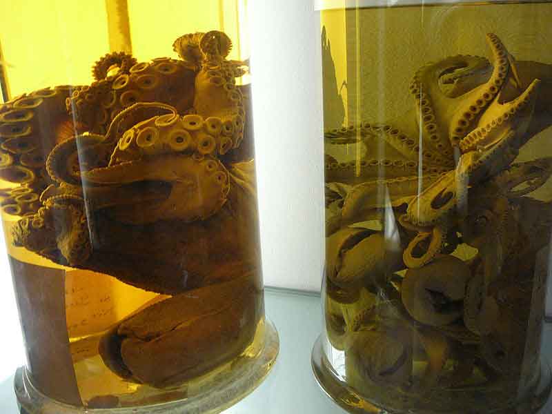
Formaldehyde is commonly used as a preservative.
Formaldehyde is a colorless, strong-smelling, flammable chemical that is produced industrially and used in building materials such as particleboard, plywood, and other pressed-wood products. In addition, it is commonly used as a fungicide, germicide, and disinfectant, and as a preservative in mortuaries and medical laboratories. Formaldehyde also occurs naturally in the environment. It is produced during the decay of plant material in the soil and during normal chemical processes in most living organisms. It is also a combustion product found in tobacco smoke.
People are exposed primarily by inhaling formaldehyde gas or vapor from the air or by absorbing liquids containing formaldehyde through the skin. Workers who produce formaldehyde or products that contain formaldehyde—as well as laboratory technicians, certain health care professionals, and mortuary employees—may be exposed to higher levels of formaldehyde than people in the general population.
The general public may be exposed to formaldehyde by breathing contaminated air from sources such as pressed-wood products, tobacco smoke, and automobile tailpipe emissions. Another potential source of exposure to formaldehyde is the use of unvented fuel-burning appliances, such as gas stoves, wood-burning stoves, and kerosene heaters.
Studies of workers exposed to high levels of formaldehyde, such as industrial workers and embalmers, have found that formaldehyde causes myeloid leukemia and rare cancers, including cancers of the paranasal sinuses, nasal cavity, and nasopharynx.
The U.S. Environmental Protection Agency recommends the use of "exterior-grade" pressed-wood products to limit formaldehyde exposure in the home. Formaldehyde levels in homes and work settings can also be reduced by ensuring adequate ventilation, moderate temperatures, and reduced humidity levels through the use of air conditioners and dehumidifiers.
EPA: Probable human carcinogen, based on limited evidence in humans, and sufficient evidence in animals. IARC: Carcinogenic to humans . NTP: Reasonably anticipated to be a human carcinogen
There is sufficient evidence in humans for the carcinogenicity of formaldehyde. Formaldehyde causes cancer of the nasopharynx and leukaemia. Also, a positive association has been observed between exposure to formaldehyde and sinonasal cancer. There is sufficient evidence in experimental animals for the carcinogenicity of formaldehyde. The Working Group was not in full agreement on the evaluation of formaldehyde causing leukaemias in humans, with a small majority viewing the evidence as sufficient of carcinogenicity and the minority viewing the evidence as limited. Particularly relevant to the discussions regarding sufficient evidence was a recent study accepted for publication which, for the first time, reported aneuploidy in blood of exposed workers characteristic of myeloid leukaemia and myelodysplastic syndromes, with supporting information suggesting a decrease in the major circulating blood-cell types and in circulating haematological precursor cells. The authors and Working Group felt that this study needed to be replicated. Formaldehyde is carcinogenic to humans (Group 1).
IARC. Monographs on the Evaluation of the Carcinogenic Risk of Chemicals to Humans. Geneva: World HealthOrganization, International Agency for Research on Cancer, 1972-PRESENT. (Multivolume work). Available at:http://monographs.iarc.fr/ENG/Classification/index.php, p. V100F 430 (2012)
Cancer Classification: Group B1 Probable Human Carcinogen
USEPA Office of Pesticide Programs, Health Effects Division, Science Information Management Branch: "ChemicalsEvaluated for Carcinogenic Potential" (April 2006)
CLASSIFICATION: B1; probable human carcinogen. BASIS FOR CLASSIFICATION: Based on limited evidence in humans, and sufficient evidence in animals. Human data include nine studies that show statistically significant associations between site-specific respiratory neoplasms and exposure to formaldehyde or formaldehyde-containing products. An increased incidence of nasal squamous cell carcinomas was observed in long-term inhalation studies in rats and in mice. The classification is supported by in vitro genotoxicity data and formaldehyde's structural relationships to other carcinogenic aldehydes such as acetaldehyde. HUMAN CARCINOGENICITY DATA: Limited. ANIMAL CARCINOGENICITY DATA: Sufficient.
U.S. Environmental Protection Agency's Integrated Risk Information System (IRIS). Summary on Formaldehyde (50-00-0). Available from, as of December 15, 2014: http://www.epa.gov/iris/
A2; Suspected human carcinogen.
American Conference of Governmental Industrial Hygienists. Threshold Limit Values for Chemical Substances andPhysical Agents and Biological Exposure Indices. ACGIH, Cincinnati, OH 2014, p. 32
Formaldehyde: Known to be a human carcinogen.
DHHS/National Toxicology Program; Thirteenth Report on Carcinogens: Formaldehyde (50-00-0) (2014). Availablefrom, as of December 12, 2014: http://ntp.niehs.nih.gov/pubhealth/roc/roc13/index.html
IARC-1, NIOSH-Ca, NTP-R, OSHA-Ca, TLV-A1, EPA-B1
HE1, HE9, HE11, HE14
The substance can be absorbed into the body by inhalation.
The substance can be absorbed into the body by inhalation, through the skin and by ingestion.
The substance can be absorbed into the body by inhalation and by ingestion.
inhalation, skin and/or eye contact
irritation eyes, nose, throat, respiratory system; lacrimation (discharge of tears); cough; wheezing; [potential occupational carcinogen]
Cough. Sore throat. Burning sensation behind the breastbone. Headache. Shortness of breath.
Cough. Sore throat. Burning sensation. Laboured breathing.
Redness.
Redness. Pain. Skin burns.
Redness. Pain.
Watering of the eyes. Redness. Pain. Blurred vision.
Watering of the eyes. Redness. Pain. Severe burns.
Redness. Pain. Burns.
Burns in mouth and throat. Nausea. Abdominal pain. Shock or collapse.
Burning sensation in the throat and chest.
Dermal (Skin), Gastrointestinal (Digestive), Immunological (Immune System), Respiratory (From the Nose to the Lungs)
Eyes, respiratory system
[nasal cancer]
| Organism | Test Type | Route | Dose | Effect | Reference |
|---|---|---|---|---|---|
| man | TDLo | oral | 643 mg/kg (643 mg/kg) | LUNGS, THORAX, OR RESPIRATION: RESPIRATORY OBSTRUCTION; GASTROINTESTINAL: ULCERATION OR BLEEDING FROM STOMACH; GASTROINTESTINAL: NAUSEA OR VOMITING | Japanese Journal of Toxicology., 4(261), 1991 |
| women | LDLo | oral | 108 mg/kg (108 mg/kg) | Practical Toxicology of Plastics, Lefaux, R., Cleveland, OH, Chemical Rubber Co., 1968, -(328), 1968 | |
| human | TCLo | inhalation | 17 mg/m3/30M (17 mg/kg) | SENSE ORGANS AND SPECIAL SENSES: LACRIMATION: EYE; LUNGS, THORAX, OR RESPIRATION: OTHER CHANGES | JAMA, Journal of the American Medical Association., 165(1908), 1957 [PMID:13480837] |
| man | TDLo | oral | 646 mg/kg (646 mg/kg) | GASTROINTESTINAL: GASTRITIS; GASTROINTESTINAL: ULCERATION OR BLEEDING FROM STOMACH; GASTROINTESTINAL: NAUSEA OR VOMITING | Japanese Journal of Toxicology., 4(261), 1991 |
| women | LDLo | oral | 1 mL/kg (1 mg/kg) | BEHAVIORAL: COMA; CARDIAC: OTHER CHANGES; GASTROINTESTINAL: ALTERATION IN GASTRIC SECRETION | Intensive Care Medicine., 23(708), 1997 |
| Resident Soil (mg/kg) | 1.7E+01 |
|---|---|
| Industrial Soil (mg/kg) | 7.3E+01 |
| Resident Air (ug/m3) | 2.2E-01 |
| Industrial Air (ug/m3) | 9.4E-01 |
| Tapwater (ug/L) | 4.3E-01 |
| Risk-based SSL (mg/kg) | 8.7E-05 |
